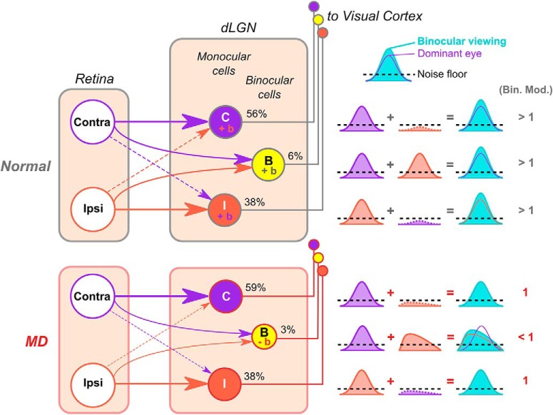
Figure 13.
Schematic model of abnormal binocular integration in mouse dLGN following long-term critical-period MD. Summary of main findings. In normal mice, binocular dLGN neurons relay binocularly matched visual inputs to V1. Monocular inputs sum to give rise to larger responses during binocular viewing (binocular facilitation, indicated by “+b” and binocular modulation values of > 1). Monocular dLGN neurons also display binocular facilitation because the input from the nondominant eye, albeit subthreshold, acts in synergy with the dominant-eye input. In MD mice, the percentage of binocular dLGN neurons is reduced, and surviving binocular neurons relay mismatched visual information to V1. MD mice lack binocular facilitation of binocular and monocular dLGN neurons, potentially due to the mismatch in suprathreshold and subthreshold visual inputs. In the case of binocular neurons, binocular viewing leads to even lower activity levels compared with monocular viewing through the dominant eye (binocular suppression, indicated by “−b” and binocular modulation values < 1). For simplicity, the model does not depict other modes of binocular modulation, such as binocular activation and complete suppression (“both only” and “suppressed” in Fig. 10).

