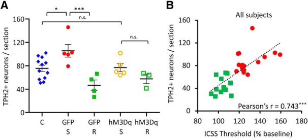
Figure 7.

Chronic activation of amygdalar CRH+ neurons abolishes stress-induced gain of TPH2 in susceptible rats. A, Quantification of TPH2+ neurons in the DRv from pooled control (C; blue diamonds, n = 12 rats), GFP-expressing susceptible (GFP-S; red solid circles, n = 5 rats), GFP-expressing resilient (GFP-R; green solid squares, n = 4 rats), hM3Dq-expressing susceptible (hM3Dq-S; orange hollow circles, n = 5 rats) and hM3Dq-expressing resilient (hM3Dq-R; green hollow squares, n = 3 rats) groups. Counts were averaged across rats and plotted as mean ± SEM. *p < 0.05, ***p < 0.001; n.s., not significant (p > 0.05). B, Correlation analysis of DRv TPH2+ neurons and ICSS thresholds (average of Days 19–21) for all stressed rats (wild-type and Crh-cre) used in this study. Rats were classified as susceptible (red solid circles; n = 16) or resilient (green solid squares; n = 14), across genotypes and virus groups. Pearson's product-moment correlation coefficient shown at the bottom right for each analysis. ***p < 0.001.
