
Fig. 1. Fate mapping as a tool to specifically label microglia/CAMs.
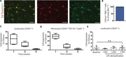
(A and B) Representative immunohistochemical images of the uninjured spinal cord demonstrated very high recombination efficiency at 4 weeks after tamoxifen (tdTom reporter, red; IBA1, green) (A). This observation was also reflected in quantification (B). (C and D) Flow quantification of reporter expression in the blood of uninjured mice demonstrated a progressive reduction in tdTom+ leukocytes (C) or monocytes (D). (E) Flow quantification of the blood of injured mice demonstrated no difference in reporter expression in CD45+ leukocytes. n = 4 (B), n = 3 to 4 (C to E). Error bars indicate ± SEM. DPI, days post-LPC injection. Scale bars, 25 μm.