Figure 4.
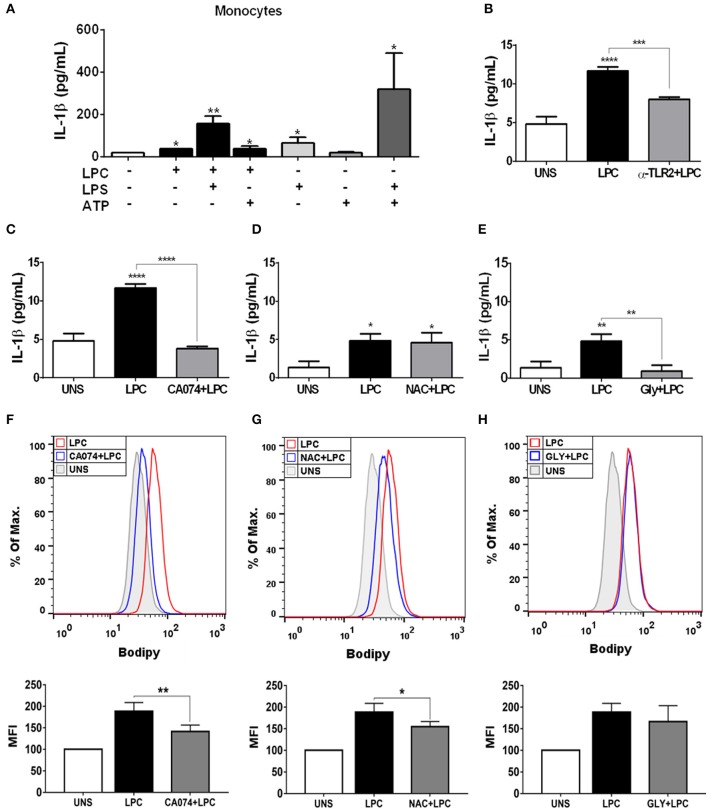
Lysophosphatidylcholine (LPC) induces IL-1β release and foam cell formation mediated by an inflammasome-dependent pathway in human monocytes. Human monocytes (A) were pretreated with LPS (500 ng/ml) 4 h before treatment with 1 μg of LPC for 24 h. After this treatment, the cells were treated with ATP (1 mM). One hour later, the supernatants were collected, and the levels of IL-1β were measured by ELISAs. Data are expressed as the average of triplicate wells. In addition, human monocytes were pretreated with different inhibitors: (B) TLR2 neutralizing antibody (α-TLR2), (C) cathepsin B inhibitor (CA-074), (D) reactive oxygen species (ROS) inhibitor [N-acetyl-l-cysteine (NAC)], and (E) inhibitor of potassium efflux [glibenclamide (GLY)] for 1 h and stimulated with 1 μg/ml of LPC for 24 h; the supernatants were collected, and the levels of IL-1β were measured by ELISAs. The bar graph represents one experiment performed twice in triplicate. Human monocytes were pretreated with different inhibitors (F) cathepsin B inhibitor (CA-074), (G) reactive oxygen species (ROS) inhibitor [N-acetyl-L-cysteine (NAC)], and (H) inhibitor of potassium efflux [glibenclamide (GLY)] for 1 h and stimulated with 1 μg/ml of LPC for 24 h, the lipid droplets were stained in cells with BODIPY and analyzed by flow cytometry. Histograms are representative of three independent experiments. Each bar graphic represents the mean fluorescence intensity (MFI), and bars show significant differences and represent the 95% confidence interval (*p < 0.05, **p < 0.01, ***p < 0.005, and ****p < 0.0001) of the cells stimulated with LPC or UNS (unstimulated cells).

