Figure 2.
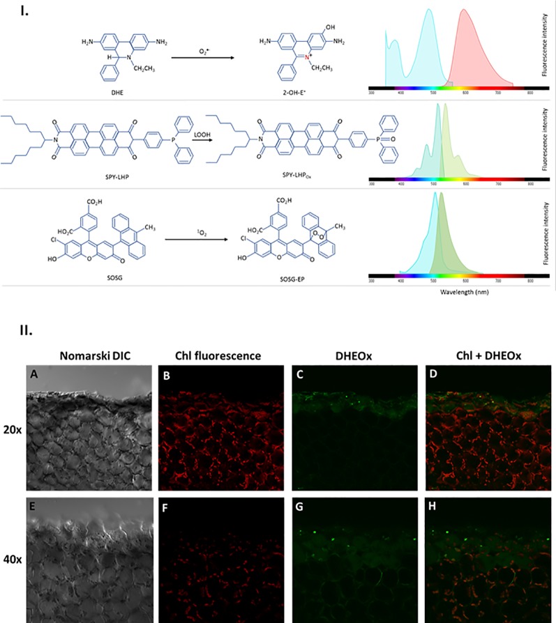
I. Principles of ROS detection and fluorochrome spectral properties (A) DHE oxidation by O2 •− forming 2-OH-E+ providing fluorescence with excitation/emission maxima of ~500/590 nm. (B) SPY-LHP oxidation by LOOH forming SPY-LHPOx providing fluorescence with excitation/emission maxima of ~524/535 nm. (C) SOSG oxidation by 1O2 forming fluorescent SOSG-EP with excitation/emission maxima of ~504/525 nm). II. Superoxide anion radical imaging in cells of WT Arabidopsis leaves detected by confocal laser scanning microscope. The panels (from left to right) represents the Nomarski DIC (A, E) chl fluorescence (B, F) DHEox fluorescence (C, G) and combined (chl fluo + DHEox) (D, H) channel following 30 min of incubation in DHE [100 μM (upper panel)/250 μM (lower panel)] in the presence of 0.01% DMSO. The margins indicate the site of mechanical injury visualized under objective of of 20× (upper panel) and 40× (lower panel). The fluorescence signal was visualized with an excitation (λex) and emission (λem) wavelengths of 488 nm and 505–605 nm respectively. Chloroplasts imaging was achieved with laser excitation at 543 nm and emission recorded at 655–755 nm.

