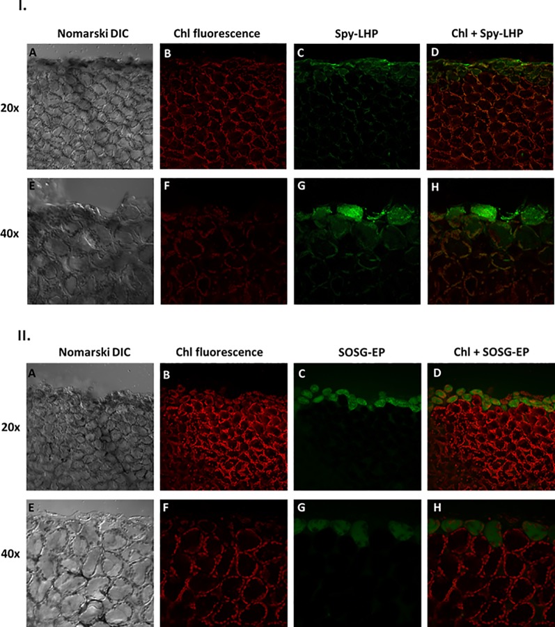
Figure 3.
I. Imaging of lipid hydroperoxide within Arabidopsis leaves by confocal microscopy. The panels (from left to right) represent DIC (A, E); chlorophyll fluorescence (B, F); Spy-LHPOx fluorescence (C, G) and combined channel (chl fluorescence + Spy-LHPOx) (D, H) following 30 min of incubation in 50 μM Spy-LHP under objective of 20× (upper panel) or 40× (lower panel). The formation of LOOH was measured in Arabidopsis leaves with excitation (λex) and emission (λem) wavelength of 488 nm and 505–550 nm, respectively. II. Confocal microscopy imaging of singlet oxygen formed during mechanical injury of Arabidopsis leaves. The panels represent (from left to right): DIC (A, E); chl fluorescence (B, F) SOSG-EP fluorescence (C, G) and combined channel (chl fluorescence + SOSG-EP) (D, H) under 20× and 40× objective following 30 min of incubation in 50 μM SOSG. The SOSG-EP fluorescence was excited by 488 nm and emission recorded at 505–525 nm.

