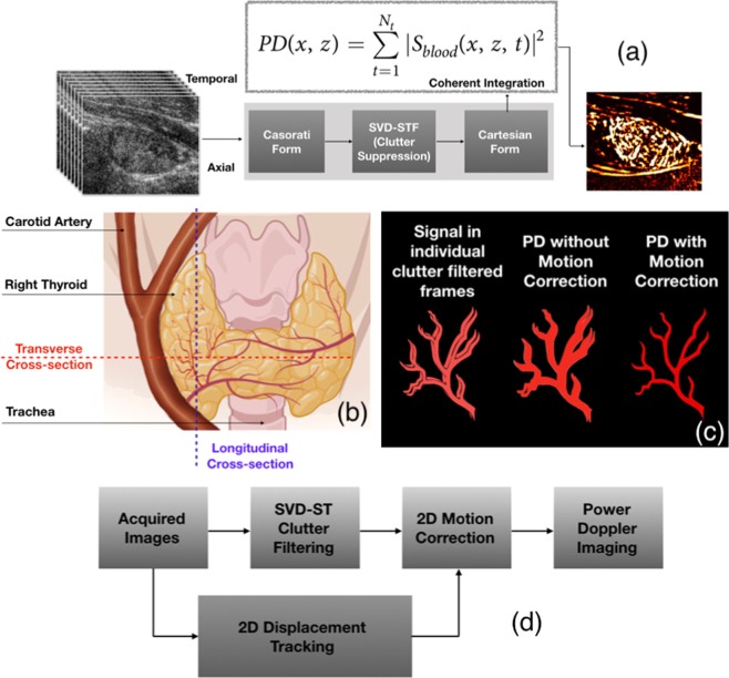
Figure 1.
An illustrative example of impact of motion on blood flow visualization in thyroid imaging, across transverse and longitudinal views. (a) Overviews the steps involved in conventional SVD based spatiotemproal clutter-filtered power Doppler imaging involving a large ensemble of ultrasound images obtained at high frame-rate. (b) Displays the anatomical position of the thyroid gland, the pulsating carotid artery and the rigid trachea, with respect to the longitudinal and transverse planes. The image in (b) was created with BioRender.com (c) illustrates the impact of motion on coherent integration of the Doppler ensemble, and its potential impact on the visualization of the blood flow signal14. (d) Outlines the steps involved in correcting in-plane motion, prior to Doppler integration for improved visualization, as demonstrated in14.

