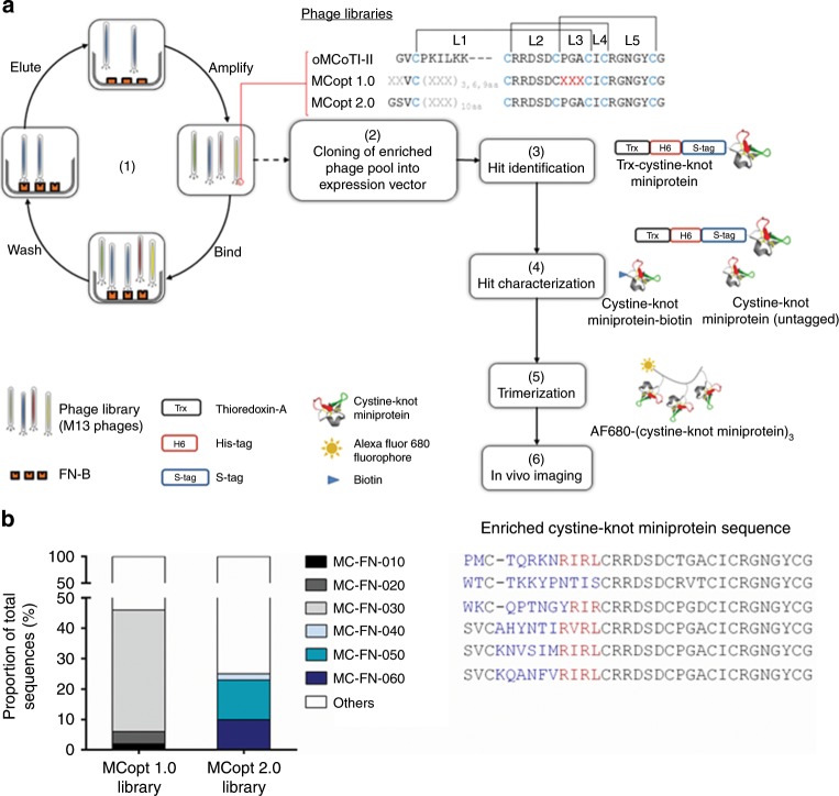
Fig. 1. Enrichment of clones with a common sequence motif by library screening against EDB.
a EDB-specific ligand selection and development of an imaging agent. (1) Three successive rounds of screening of MCopt 1.0 and MCopt 2.0 phage libraries (both based on the oMCoTI-II sequence framework) were performed against a hexahistidine (H6)-tagged single EDB-domain (FN-B) fragment. Disulfide bonds (brackets) between cysteine residues (blue), randomized positions for any random amino acid except cysteine (X in gray), and amino acid substitutions to 50% (X in red) are indicated. L1 to L5 represent the loop positions. (2) Cystine-knot miniprotein sequences were cloned into expression vector for Trx-cystine-knot miniprotein production. (3) Hit identification of individual clones was performed by ELISA-based binding analysis (Trx-cystine-knot miniprotein), determination of expression rate, and sequencing. (4) Hits were characterized with regard to affinity (with untagged cystine-knot miniprotein), specificity (Trx-cystine-knot miniprotein, cystine-knot miniprotein-biotin), and functionality (Trx-cystine-knot miniprotein). (5) Trimerization of lead cystine-knot miniprotein candidates and Alexa Fluor 680 fluorophore conjugation was performed to allow (6) imaging of tumor vasculature in vivo in a mouse model xenografted with a human glioblastoma cell line. b Enrichment of cystine-knot miniprotein sequences after three screening rounds of phage display libraries MCopt 1.0 and MCopt 2.0. Variable amino acids (blue letters) and the common R-I/V-R-(L) motif (red) are indicated.

