Figure 1.
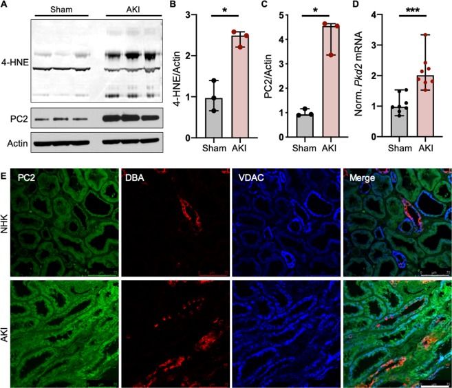
PC2 levels are increased in pathologically stressed kidneys. (A) Normal (Sham) and AKI-afflicted mouse kidneys were immunoblotted for 4-HNE and PC2. Each band represents one biological replicate. Full-length blots shown in Fig. S6. (B,C) Quantification of 4-HNE and PC2 protein abundance in Sham and AKI kidneys, normalized to actin. *p < 0.05 as determined by Mann Whitney U test. Data presented as median with range. Sample size n = 3 biological replicates per group. (D) Normalized mRNA expression of Pkd2 in Sham and AKI-afflicted mouse kidneys. Gapdh used as internal control. Sample size n = 8 biological replicates per group. ***p < 0.001 as determined by Mann Whitney U test. Data presented as median with range. (E) Normal human kidneys (NHK) or kidneys with acute kidney injury (AKI) were stained for PC2 (green), DBA (red), a marker for collecting ducts, and VDAC (blue), an outer mitochondrial membrane protein. Scale bar, 75 μm.

