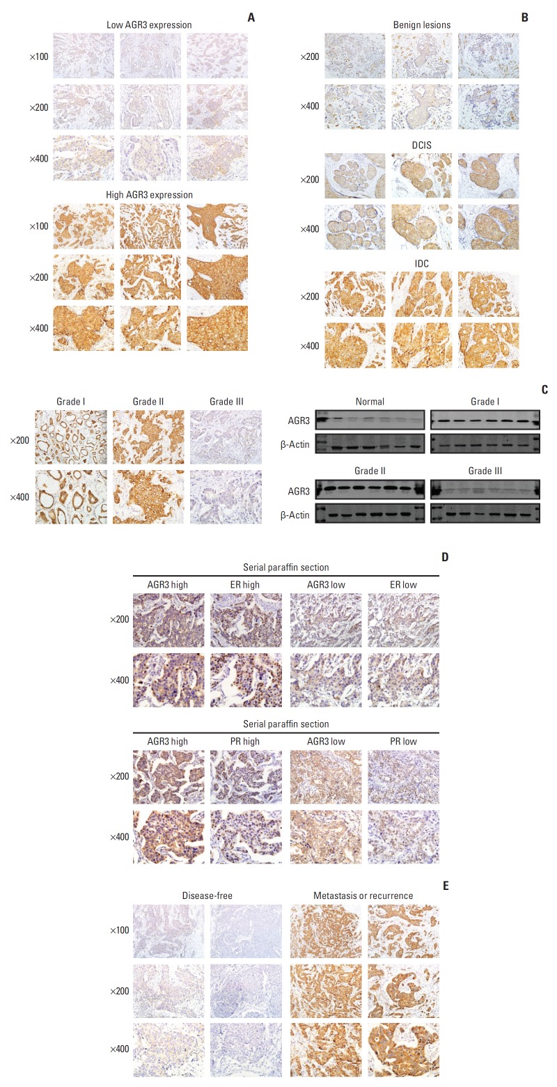
Fig. 1.
Anterior gradient 3 (AGR3) expression increased with the development of breast tumor malignancy and was negatively correlated with histological grade but positively correlated with estrogen receptor (ER)/progesterone receptor (PR) status. (A) Representative immunohistochemistry (IHC) images of AGR3 low expression group (upper) and AGR3 high expression group (lower). (B) AGR3 expression increased with progression of malignancy degree of lesions. AGR3 expression of benign lesions was low, ductal carcinoma in situ (DCIS) was higher and IDC was the highest. (C) Representative IHC images of AGR3 expression in different histological grades of IDC (left panel). Western blot analysis of AGR3 expression in the frozen breast tumor specimens consisting of normal tissues, grade I, grade II, and grade III. Every type of tissues had 6 cases. β-actin was used as a loading control (right panel). (D) The expression of AGR3, ER, and PR were detected by IHC in serial paraffin sections. The upper panel was representative IHC images of AGR3 and ER. The lower was AGR3 and PR. (E) Representative IHC images of AGR3 expression in disease-free group and recurrence or metastasis group.

