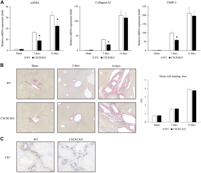
Fig. 2.
Role of CXC chemokine receptor 2 (CXCR2) on biliary fibrosis after bile duct ligation (BDL). A: mRNA expression of profibrotic genes in the liver after BDL. α-Smooth muscle actin (α-SMA), collagen 1α1, and tissue inhibitor of metalloproteinase-1 (TIMP-1) expression was significantly less in CXCR2−/− [knockout (KO)] mice than wild-type (WT) mice at 3 days. α-SMA expression was significantly less in CXCR2−/− mice than wild-type mice at 14 days. KO, knockout. B: Sirius Red staining. There was no difference in biliary fibrosis between wild-type mice and CXCR2−/− mice after BDL. Quantitative morphometric analysis of Sirius Red staining showed no significant difference in fibrosis between wild-type mice and CXCR2−/− mice. Original magnification: ×100. Data are means ± SE with n = 3–9 per group. Student’s t test was used for the statistical analysis. *P < 0.05, compared with wild type. C: CK7 staining. There was no difference in ductular reaction between wild-type mice and CXCR2−/− mice at 14 days after BDL.

