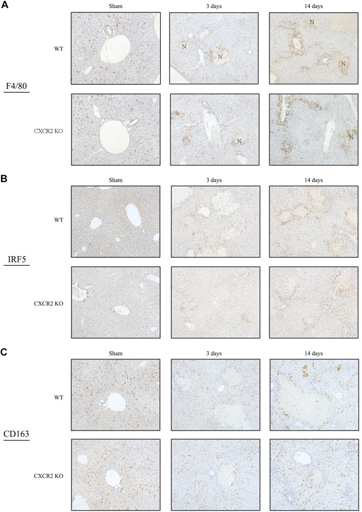
Fig. 4.
Macrophage infiltration after bile duct ligation (BDL). A: F4/80 immunohistochemical staining. Macrophages accumulated to the necrotic area and periportal area in both wild-type (WT) mice and CXC chemokine receptor 2 knockout (CXCR2−/−; CXCR2KO) mice. Original magnification: ×200. B: macrophage polarization after BDL. IRF5 immunohistochemical staining showed that M1 macrophages preferably accumulated to the injured area in wild type. C: CD163 immunohistochemical staining showed that M2 macrophages existed in the sinusoid throughout liver tissue in wild type. CXCR2−/− mice also had similar pattern of the accumulation of M1 and M2 macrophages. Original magnification: ×200.

