Fig. 4.
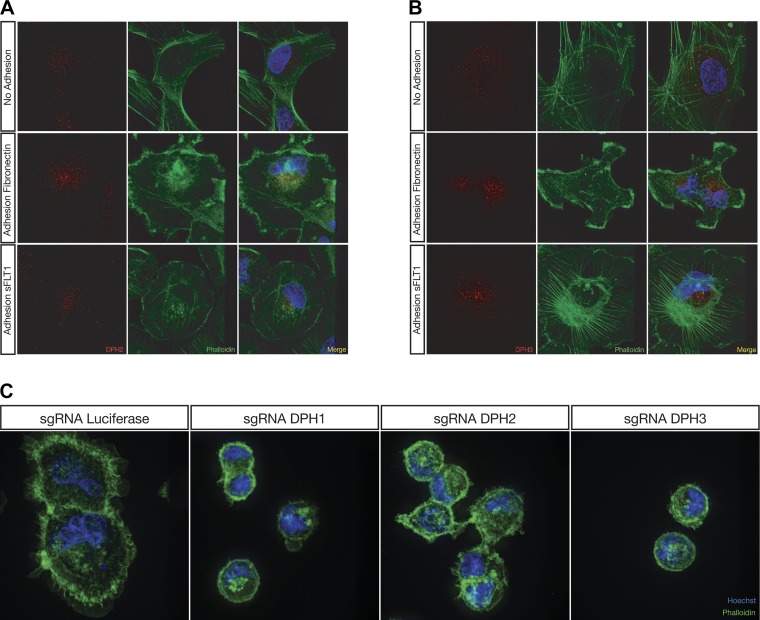
Diphthamide biosynthesis genes localize to a distinct subcellular compartment during adhesion, whereas lentiCRISPR-mediated knockout of diphthamide biosynthesis genes in human podocytes results in a morphological phenotype when adhering to fibronectin. The red channel represents either diphthamide biosynthesis protein (DPH)2 (A) or DPH3 (B), the green channel represents actin staining with phalloidin, and the blue channel is nuclear staining with Hoechst 33342. Podocytes grown on glass coverslips and serum starved overnight exhibited a diffuse cytoplasmic localization of DPH2 (A) and DPH3 (B) protein. Upon adhesion to either fibronectin or soluble Fms-like tyrosine kinase-1 (sFLT1)/Fc, DPH2 (A) and DPH3 (B) protein localized to a perinuclear compartment. Pod-Cas9 cells expressing single guide RNAs (sgRNAs) against DPH1, DPH2, and DPH3 exhibited a spreading defect when plated on fibronectin compared with luciferase sgRNA (C).

