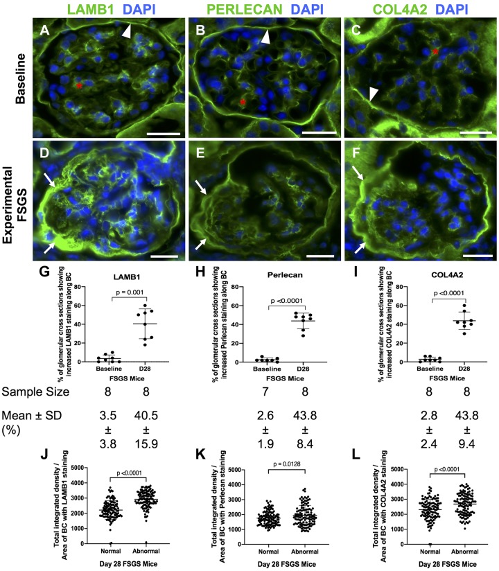
Fig. 1.
Laminin-β1 (LAMB1), perlecan, and collagen type IV-α2 (COL4A2) staining increase in experimental focal segmental glomerulosclerosis (FSGS). A−C: a normal mouse at baseline. Staining (green) for LAMB1 (A), perlecan (B), and COL4A2 (C) was present in parietal epithelial cell (PECs) in mice at baseline. They were detected along Bowman’s capsule (BC; white arrowheads) and in the mesangial matrix (red asterisk) but not along the glomerular basement membrane. Nuclei were stained with DAPI (blue). D−F: FSGS day 28. Staining increased for LAMB1 (D), perlecan (E), and COL4A2 (F) in mice at FSGS day 28 in a PEC distribution in FSGS (white arrows show examples). G−I: graphs of quantification of glomerular cross sections. The percentage of glomerular cross sections with increased staining along BC was increased for LAMB1 (G), perlecan (H), and COL4A2 (I) staining at day 28 FSGS compared with baseline. The number of mice quantitated is shown below each graph. J−L: graphs of quantification of fluorescence intensity. Quantification of fluorescence intensity showed a significant increase in the intensity of LAMB1 (J), perlecan (K), and COL4A2 (L) staining in abnormal glomeruli in FSGS. Original magnification: ×400. Scale bars = 20 μm.

