Fig. 3.
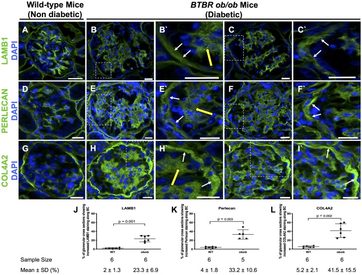
Laminin-β1 (LAMB1), perlecan, and collagen type IV-α2 (COL4A2) increased in diabetic ob/ob mice at 24 wk. A, D, and G: nondiabetic wild-type (WT) mice. LAMB1 (A), perlecan (D), and COL4A2 (G) staining (green) was detected along Bowman’s capsule (BC) in nondiabetic WT mice. Nuclei were stained with DAPI (blue). B, C, E, F, H, and I: representative staining in two diabetic ob/ob mouse glomeruli for LAMB1 (B and C), perlecan (E and F), and COL4A2 (H and I) was increased along BC. The insets at higher magnification show increased staining along BC (white arrows) and the mesangium (yellow arrows) for LAMB1 (B′ and C′), perlecan (E′ and F′), and COL4A2 (H′ and I′). J−L: quantification. The percentage of glomerular cross-sections with increased LAMB1 (J), perlecan (K), and COL4A2 (L) along BC was significantly higher in diabetic ob/ob mice than in nondiabetic WT mice. Original magnification: ×400. Scale bars = 20 μm.

