Fig. 9.
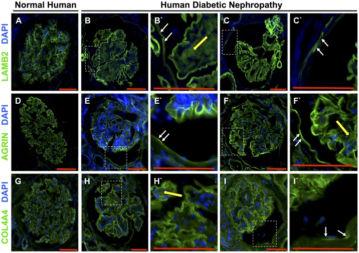
Laminin-β2 (LAMB2), agrin, and collagen type IV-α4 (COL4A4) staining in human diabetic nephropathy. A–C: LAMB2 staining. A: LAMB2 staining was restricted to the glomerular basement membrane (GBM) in the normal glomerulus. B and C: LAMB2 was detected along Bowman’s capsule in glomeruli from two different patients with diabetic nephropathy. B′ and C′: higher magnification of the insets showing de novo staining for LAMB2 along Bowman’s capsule (white arrows) and in the mesangium (yellow arrow). D–F: agrin staining. D: agrin staining localized to the GBM in the normal glomerulus. E and F: agrin was also detected along Bowman’s capsule in glomeruli from two different patients. E′ and F′: higher magnification of the insets showing staining for agrin along Bowman’s capsule (white arrows) and in the mesangium (yellow arrow). G–I: COL4A4 staining. G: COL4A4 staining localized to the GBM in the normal glomerulus. H and I: COL4A4 was also detected along Bowman’s capsule in glomeruli from two different patients with diabetic nephropathy. H′ and I′: higher magnification showing COL4A4 staining along Bowman’s capsule (white arrows) and in the mesangium (yellow arrow). Original magnification: ×200. Scale bars = 100 μm.

