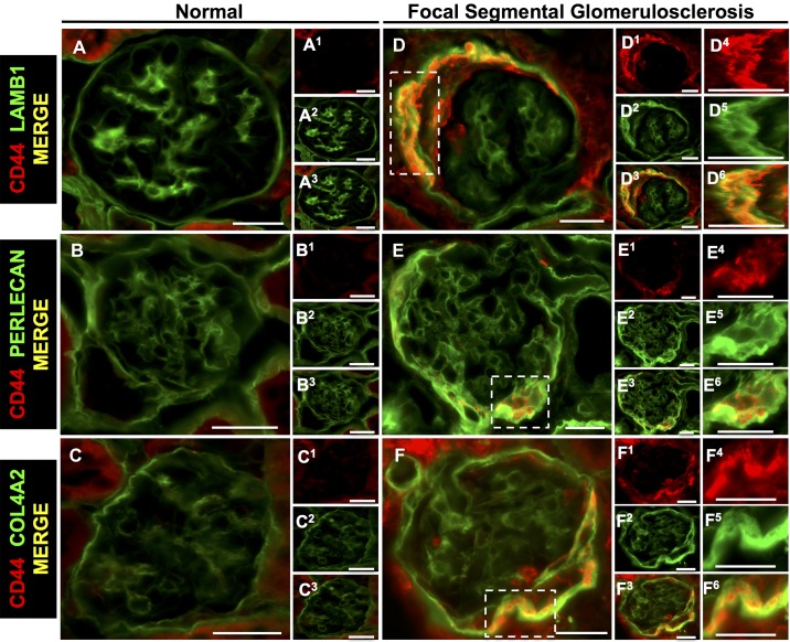
Fig. 10.
Parietal epithelial cell (PEC)-derived proteins colocalize with the activated PEC marker CD44 in mice with experimental focal segmental glomerulosclerosis (FSGS). Double staining was performed for CD44 (red) and PEC-derived extracellular matrix proteins (green). A−C: normal mice. The merged images show staining for LAMB1 (A), perlecan (B), and COL4A2 (C) but not CD44 in normal glomeruli. A1−C3: individual stains for LAMB1, perlecan, and COL4A2. D−F: FSGS mice. Merged images show colocalization (yellow) of CD44 in PECs with LAMB1 (D and D3), perlecan (E and E3), and COL4A2 (F and F3). D1, D2, E1, E2, F1, and F2: corresponding individual stainings for LAMB1 (D1 and D2), perlecan (E1 and E2), and COL4A2 (F1 and F2). Single and merged higher magnifications of the insets are shown in D4–F6. These results are consistent with activated PECs expressing increased LAMB1, perlecan, and COL4A2 isoforms in FSGS. Original magnification: ×400. Scale bars = 20 μm.

