Fig. 3.
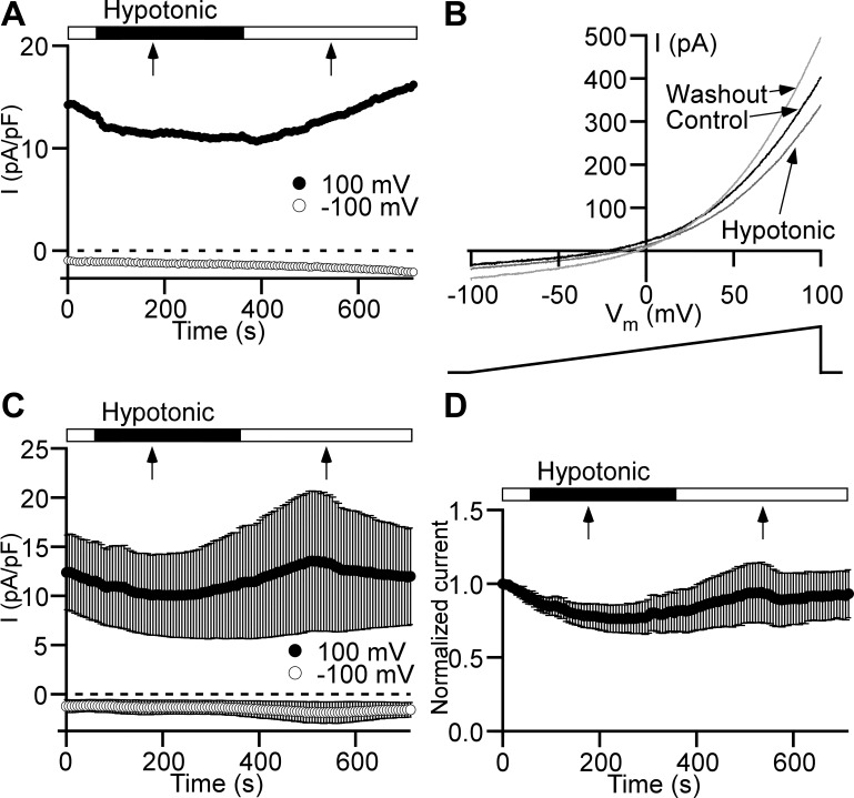
Low osmotic pressure does not cause Cl− current increase in detrusor smooth muscle cells. A: time course of Cl− current (I) measured with a ramp protocol at +100 mV (●) and −100 mV (○). Hypotonic solution (182 mOsm) was applied via a global perfusion system. The flow started at the beginning of the closed horizontal bar. The flow stopped at the time point indicated by a black arrow. The hypotonic solution application was switched back to the normotonic solution starting at the beginning of the following open horizontal bar. The flow stopped at the time moment indicated by a black arrow. B: current-membrane potential (Vm) relationships obtained from the ramp time course protocol shown in A at the following time points: immediately before starting global perfusion with hypotonic solution (Control, black trace), at the end of the 5-min hypotonic solution application (Hypotonic, dark gray trace), and 5 min after switching back to the normotonic solution (Washout, light gray trace). Arrows link traces with conditions under which they were recorded. Ramp voltage stimulus from −100 to +100 mV and 1-s duration is shown at bottom. C: Cl− current time courses (n = 6 cells, N = 4 guinea pigs) measured at +100 mV (●) and −100 mV (○). All other symbols and marks have the same meaning as in A. D: time course normalized to the first time point of the Cl− current measured at +100 mV shows no statistically significant effect of hypotonic solution (n = 6, N = 4, P > 0.05, paired Student’s t test). All other symbols and marks have the identical meaning as in A and C.

