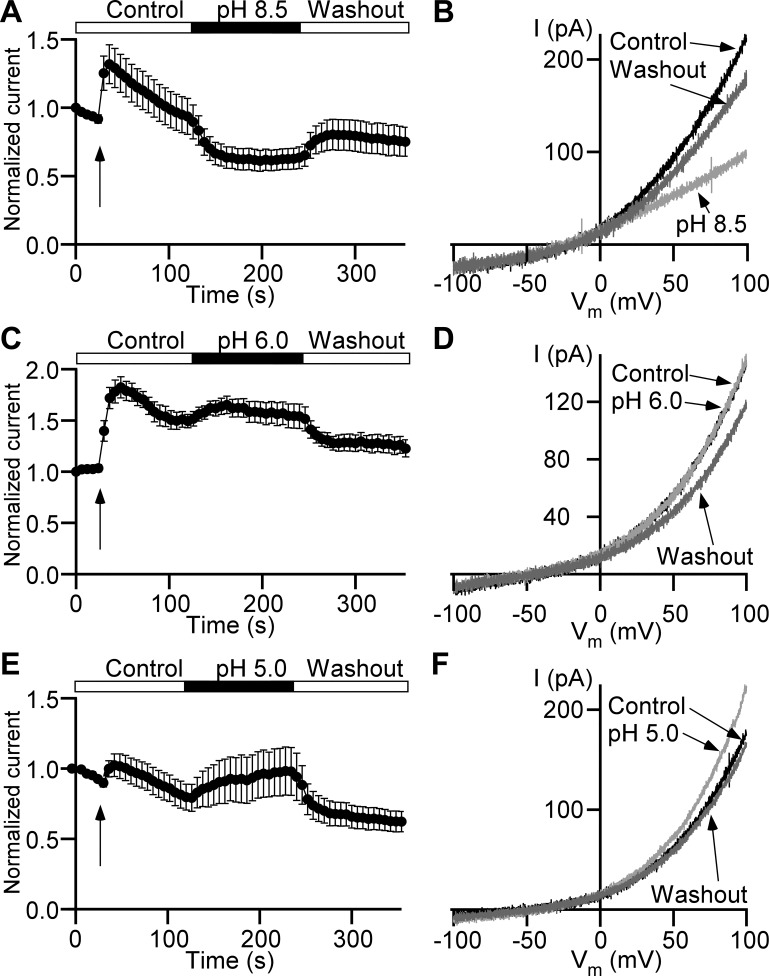
Fig. 4.
Extracellular pH modulates Cl− current in detrusor smooth muscle cells. A, C, and E: time courses of Cl− currents normalized to their first data points measured at +100 mV. Arrows indicate time points of local perfusion start. The recording begins in control bath solution with pH 7.4 followed by the isochronic application of a solution with pH 8.5 (n = 11 cells, N = 3 guinea pigs; A), pH 6.0 (n = 8, N = 3; C), or pH 5.0 (n = 11, N = 3; E) and return to pH 7.4 (Washout). B, D, and F: representative current (I) recordings evoked by ramps at time points of control (Control, black trace) right before switching to the bath solution with pH 8.5 (B), pH 6.0 (D), or pH 5.0 (F) at the end of modified pH bath solution applications (light gray trace), and same as pH 8.5 (B), pH 6.0 (D), or pH 5.0 (F) application long-duration washout (Washout, dark gray trace). Vm, membrane potential.

