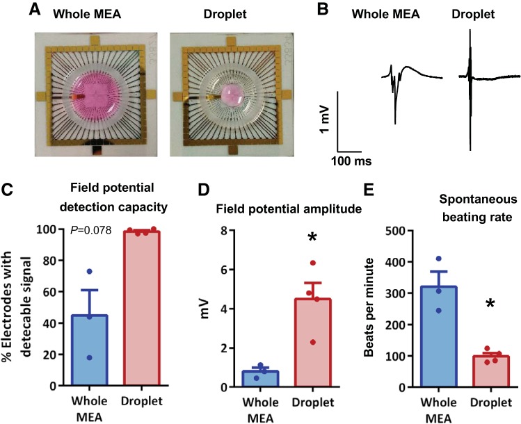
Fig. 2.
Optimal cell seeding conditions for measuring cultured neonatal rat ventricular myocyte (NRVM) field potentials. Exemplar images of NRVM seeding across the entire microelectrode array (MEA) or as a droplet over the central recording zone of the electrode array (A), with respective exemplar field potential traces (B) and percentage of electrodes detecting field potentials (C). Mean field potential amplitude (D) and spontaneous beating rate from MEAs (E). All data were acquired from mixed-sex NRVMs, immediately after a 10-min stabilization period. *P < 0.05, unpaired t tests, N = 3–4 cultures, n = 3–10 MEAs.

