Fig. 5.
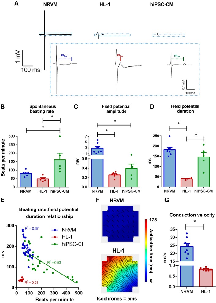
Basal electrophysiology differs among female cardiomyocyte cultures of different origin. A: exemplar field potential traces from neonatal rat ventricular myocytes (NRVMs), HL-1, and human induced pluripotent stem cell-derived cardiomyocytes (hiPSC-CMs) at full scale and cropped (at the dotted lines) to highlight repolarization. Mean spontaneous beating rate (B), field potential amplitude (C), and field potential duration (D) from all 3 cell types. E: spontaneous beating rate-field potential duration relationship was examined on a single microelectrode array (MEA) basis among NRVM, HL-1, and hiPSC-CMs. Representative activation maps (F) and mean conduction velocity (G) in NRVM and HL-1 monolayers. *P < 0.05, unpaired t tests or 1-way ANOVA with Holm-Sidak’s multiple-comparison tests. NRVM: N = 8 cultures, n = 19 MEAs; HL-1: N = 4–6 passages, n = 7–11 MEAs; hiPSC-CMs: N = 5 (3 separate cultures, 5 plating runs), n = 22 MEAs.

