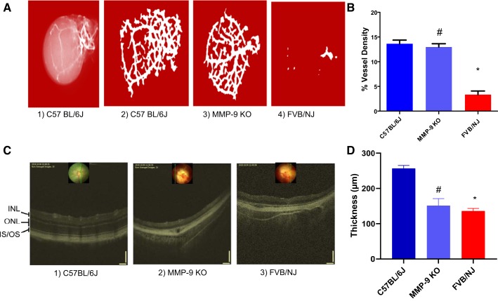
Fig. 2.
Barium sulfate angiography and the optical coherence tomographic (OCT) imaging of the mice eyes. Barium sulfate images showing the retinal microvasculature changes (A) and respective quantification of their vessels’ density (B). Absence of MMP-9 prevents the degeneration of the retinal vessels (A3). Almost complete absence of vascularization is visible in FVB/NJ mice (A4, and C3) as compared with C57BL/6J (A2, C1) and MMP-9 KO (A3, C2), confirming retinal degeneration. A progressive retinal degeneration is the hallmark in the FVB/NJ mouse as shown in the quantitative vessel density analysis (B). Also, the degeneration and thinning of the various retinal layers were successfully captured via OCT analysis along with demonstration of retinal layers’ separation/distortion occurring more in the FVB/NJ strain than in C57BL/6J, and MMP-9 KO in the back of the eyes. Although outer retinal layers run into each other in both MMP-9 KO and FVB/NJ retinae, disruption of the retinal architecture is more prominent, along with a significant loss of vasculature, in the FVB/NJ strain. Also, the layers are more severely disrupted/altered in FVB/NJ than the MMP-9 KO and C57BL/6J strains (C). Estimation of the total thickness of the retinal layers is depicted. *P < 0.0001 when compared with C57BL/6J, #P < 0.05 when compared with FVB/NJ (D). INL, inner nuclear layer; ONL, outer nuclear layer; IS, inner segment; OS, outer segment.

