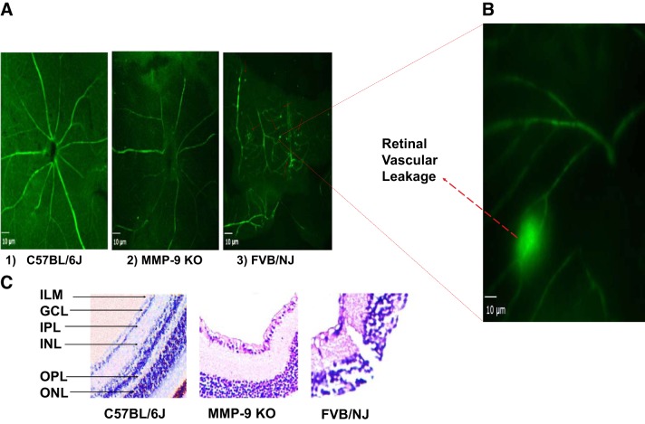
Fig. 3.
Ex vivo visualization of the BSA-FITC in retinal vessels and the architecture of inner retina in mice: representative micrographs depicting vascular integrity of the retina. The permeability or leakage in FVB/NJ is evident as shown by red arrows in A3 (and in an enlarged image shown in B) as observed on retinal flat-mount compared with C57BL/6J in A1, and MMP-9 KO in A2. Micrographs were prepared 1 h post-BSA-FITC injection through the tail vein in each strain of the mouse, scale bar = 10 μm. On further probing employing hematoxylin-eosin staining, the inner retinal layers show altered anatomy as a result of degenerative changes in FVB/NJ mice compared with MMP-9 KO and C57BL/6J. ILM, inner limiting membrane; GCL, ganglion cell layer; IPL, inner plexiform layer; INL, inner nuclear layer; OPL, outer plexiform layer; and, ONL, outer nuclear layer (C, ×200).

