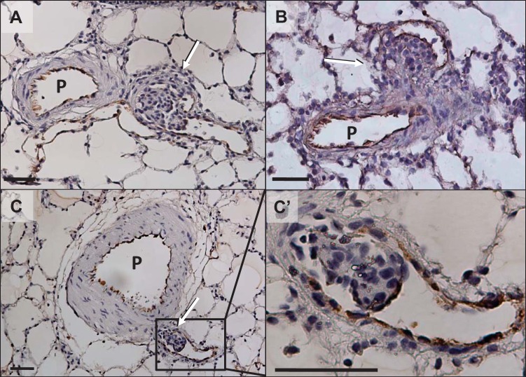
Fig. 3.
Representative photomicrographs of von Willebrand factor-stained aneurysm-type plexiform lesions in late-stage (13-wk time point) pulmonary arterial hypertension (PAH) rat lungs. A and B: typical mature lesions. C: small cellular lesion growing inside a thin-walled small branch artery with almost all plexiform cellular features except for the typical slit-like channel formation (plexiform-like lesion). C’: magnified area outlined in C. P, parent pulmonary artery; arrows indicate the complex lesion. Scale bars, 50 μm.

