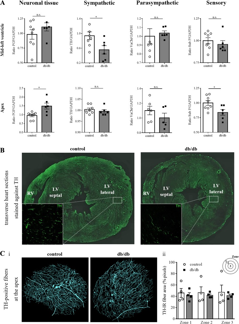
Fig. 6.
Characteristics of the cardiac autonomic innervation. A: Western blot analysis (control: n = 8, db/db: n = 6) after normalization with GAPDH confirms a heterogeneous expression of tyrosine hydroxylase (TH) in db/db mice in the midleft ventricle but not in the apex. Differences in the expression of a pan-neuronal marker (PGP 9.5) and a sensory marker (Substance P) were observed in the apex. B: transverse heart sections reveal abundant TH-positive fibers at the midleft ventricle in control hearts, whereas only sparse TH-positive fibers are present in db/db hearts, demonstrating a reduced sympathetic innervation. C: antibody staining against TH at the cardiac apex (n = 4 for control and db/db), the most peripheral area of the heart, revealed no differences in the amount of TH-positive fibers in a semiquantitative analysis. *P ≤ 0.05. LV, left ventricle; n.s., not significant; RV, right ventricle; TH-IR, TH-immunoreactivity; VAChT, vesicular acetylcholine transporter.

