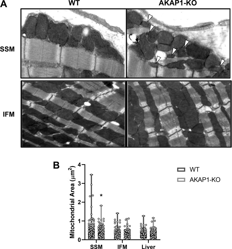
Fig. 7.
Subsarcolemmal mitochondria (SSM) ultrastructure is perturbed in A-kinase anchoring protein 1 knockout (AKAP1-KO) mice. A: image analysis from transmission electron microscopy (TEM) performed on untreated mouse heart tissues from either wild-type (WT) or AKAP1-KO mice. Arrowheads indicate areas of decreased mitochondrial area. B: quantification of mitochondrial area (μm2) for heart SSM, heart interfibrillar mitochondria (IFM), and liver mitochondria, comparing WT and AKAP1-KO mice (n = 84–124 technical replicates summed over 10 biological replicates). Data are presented as means ± SE. *P < 0.05, compared with WT.

