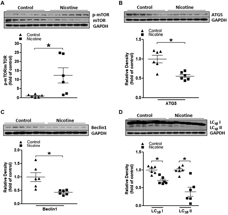
Fig. 2.
Perinatal nicotine exposure altered mammalian target of rapamycin (mTOR)/autophagy biomarker expression patterns in neonatal rat brain. Brain tissues were isolated from 9-day-old (P9) neonatal rat pups in both nicotine-exposed and saline control groups. Protein abundance in brain tissue was determined by Western blot analysis. GAPDH protein density served as internal normalized control. A: the ratio of phosphorylated (p-)mTOR to mTOR protein density is shown in the saline control (▲) and nicotine-exposed (■) groups. Protein densities of the autophagy biomarkers ATG5 (B), Beclin-1 (C), and LC3βI/II (D) are shown in saline control (▲) and nicotine-exposed (■) groups. Data are means ± SE of animals (n = 6) from each group. *P < 0.05 vs. control, as determined by Student’s t test.

