
Figure 2.
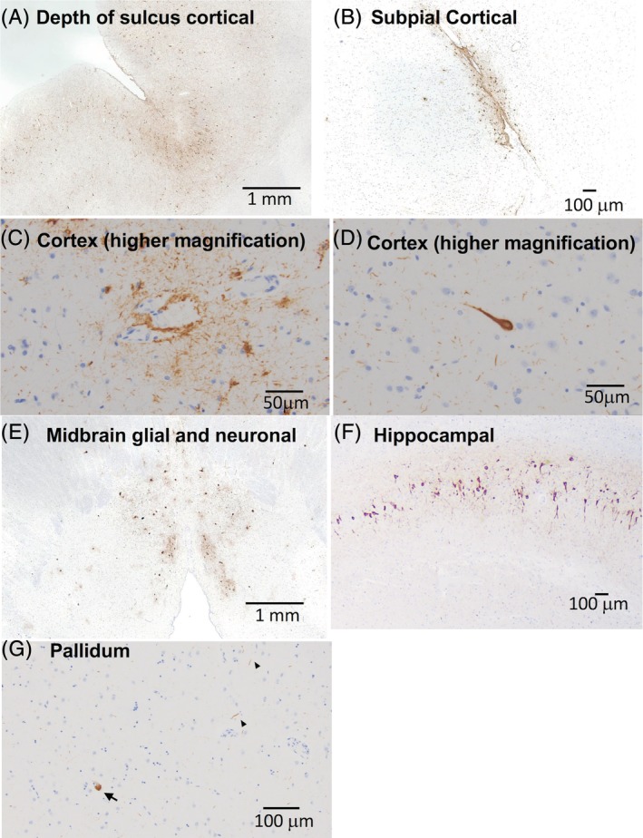
Microscopic neuropathology in a patient with ADCY5‐dyskinesia. PHF‐tau immunostained slides demonstrate patchy, but widespread, pTau pathology. (A) Sections of neocortex demonstrate neurofibrillary tangles along gyri as well as at the depths of sulci. (B) Cortical tau also consisted of subpial, astroglial tau inclusions. (C) Perivascular glial and neuritic tau pathology in the cortex. (D) High magnification of a neurofibrillary tangle. (E) Section of midbrain shows glial and neuronal tau in periaqueductal gray matter. (F) Section of hippocampus demonstrates neurofibrillary tangles in the CA1 subfield and (G) scattered tau neurites (arrowheads) and pretangles (arrow) of uncertain significance in the pallidum.