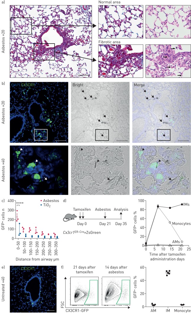
FIGURE 2.
Recruitment of monocyte-derived alveolar macrophages (Mo-AMs) is spatially restricted to areas near asbestos fibres. DAPI: 4′,6-diamidino-2-phenylindole; GFP: green fluorescent protein; IM: interstitial macrophage; FSC: forward scatter. a) The intratracheal administration of asbestos fibres induces fibrosis near bronchoalveolar duct junctions where asbestos fibres lodge. Left panel: low-power image of a medium-sized airway (Mason's trichrome). Scale bar: 100 µm. Right panels: high-power images (Mason's trichrome and haematoxylin/eosin, respectively). Areas of fibrosis develop adjacent to the airway in which asbestos fibres can be observed (arrows, bottom right panel). In contrast, alveolar structures in the distal lung parenchyma are relatively preserved. b) Top panels: representative lung histology from Cx3cr1ER-Cre×ZsGreen mice treated with tamoxifen on day 14 and 15 (10 mg via oral gavage) and harvested 21 days after asbestos exposure. Scale bar: 100 µm. Bottom row panels correspond to areas outlined in the boxes. Left panels: monocyte-derived cells are GFP+; nuclei stained with DAPI. Middle panels: phase contrast images; asbestos fibres are indicated by arrows. Right panels: merge. Bottom panels: asbestos fibres are surrounded by GFP+ cells (short arrows) and GFP− cells (arrow) with macrophage morphology. c) Quantification of GFP+ Mo-AMs in peribronchial regions in asbestos- and TiO2-treated mice. Two-way ANOVA with Tukey's multiple comparisons test. ****: p<0.0001. d) Schematic of the experimental design, and kinetics of GFP+ monocytes, tissue-resident IMs and tissue-resident AMs after tamoxifen pulse in naive Cx3cr1ER-Cre×ZsGreen mice. Percentage of GFP+ cells was assessed by flow cytometry. e) Representative fluorescent image showing GFP+ tissue-resident IMs 21 days after tamoxifen pulse in naive animals. f) Representative contour plots showing GFP expression gated on AMs from Cx3cr1ER-Cre×ZsGreen mice 21 days after tamoxifen and 14 days after asbestos instillation. Percentage of GFP+ classical monocytes, IMs and AMs was assessed by flow cytometry. All data are presented as mean±sem. n=3–5 mice per group or time-point.

