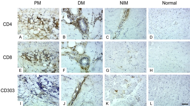
Figure 2.

Inflammatory marker staining. In PM (A, E, I), inflammatory cells were mainly located in clusters in the endomysium, whereas in DM (B, F, J), they were mostly perivascular and perifascicular. In NIMs (C, G, K), scattered inflammatory cells were present mainly in the endomysium and perimysium. There were no inflammatory cells in the normal control group (D, H, L). (Original magnification ×200, Scale bar = 300 μm).
