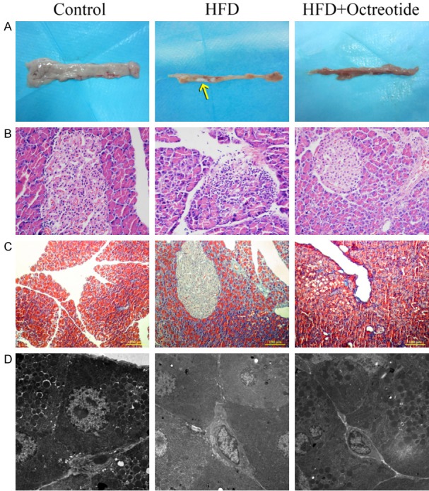
Figure 2.

Pancreatic morphology of each group. Pancreatic tissues of the HFD group became harder and smaller, and pancreatic calcification was observed (A, yellow arrow). Hematoxylin and eosin and Masson-Trichrome staining presented pancreatic acinar inflammatory infiltration and atrophy in the HFD group, and pancreatic interlobular fibers also increased in obese rats (B and C, magnification, ×200). Pancreatic ultramicrostructure was visualized by TEM: red arrow, zymogen granules; white arrow, rough endoplasmic reticula. TEM showed reduced zymogen granules, dilated rough endoplasmic reticula in the acinar cells in the HFD group, and octreotide treatment increased zymogen granules and the dilation degree of rough endoplasmic reticula was alleviated (D, magnification, ×0.7). HFD, high-fat diet; TEM, transmission electron microscopy.
