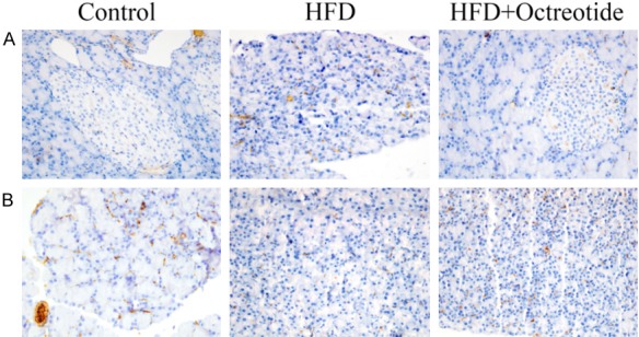
Figure 3.

Immunohistochemical staining for α-SMA and desmin. (A) α-SMA and (B) desmin were primarily localized in the periacinar regions of the pancreas. The expression of α-SMA was increased and desmin was decreased in the HFD group. Following octreotide treatment, α-SMA protein significantly decreased and desmin protein increased (magnification, ×400). α-SMA, α-smooth muscle actin; HFD, high-fat diet.
