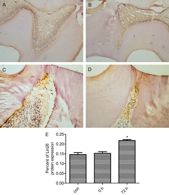
Figure 3.

Immunolocalization of the expression of Lin28 protein by 0 h and 72 h and in the control teeth after cavity preparation. (A, B) Weak immunoreactivity expression of Lin28 protein was observed by 0 h (B, ×100) and in the control teeth (A, ×100). (C, D) A higher level of activity of Lin28 was observed in the odontoblasts and the pulp cells adjacent to the cavity by 72 h (C, ×100; D, ×200). (E) The percent of Lin28 protein expression by 0 h, 72 h and in the control teeth after cavity preparation. *P < 0.05 vs the normal dentin-dental pulp. The data are presented as the mean ± SEM.
