
Figure 2.
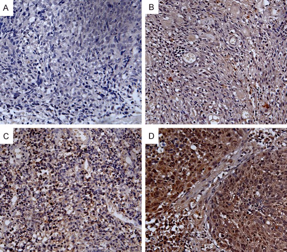
Expression analysis of ZIC2 protein as determined by immunohistochemistry (200×). ZIC2 expression was localized in the nuclei and cytoplasms of NPC cells. A. Negative staining of ZIC2 in NPC tissues. B. “+” (score 1-4, weakly positive) expression of ZIC2 in NPC tissues. C. “++” (score 5-8, positive) expression of ZIC2 in NPC tissues. D. “+++” (score 9-12, strongly positive) expression of ZIC2 in NPC tissues.