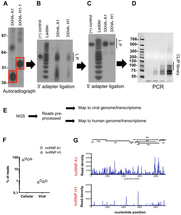
Figure 2-. CLIP with hnRNP A1 and hnRNP H1 proteins.
HEK293T cells were transfected with plasmids expressing 3xHA-tagged hnRNP A1 and hnRNP H1 (long and short isoforms) proteins. (A) Immunoprecipitated and end-labeled protein-RNA adducts were separated by SDS-PAGE, transferred to nitrocellulose membranes and visualized by autoradiography as detailed in the protocol. (B) Separation of 3’ adapter ligation products on a 15% TBE-Urea gel is shown. (C) Separation of 5’ adapter ligation products on a 15% TBE-Urea gel is shown. (D) Separation of PCR products (cycles 6, 9, 12 and 15) on a 6% TBE-Urea gel is shown.

