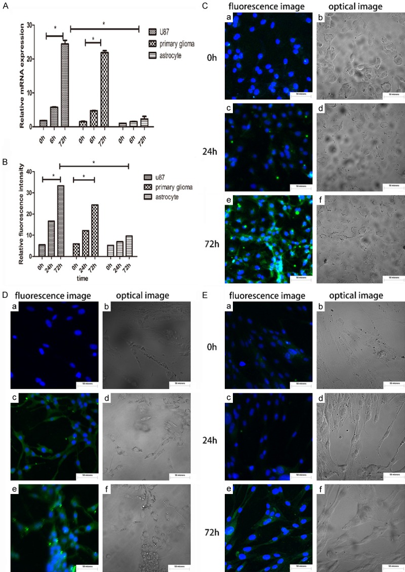
Figure 2.

(A) mRNA expression of PD-L1 at 6 h and 72 h after infection with HCMV in U87, primary glioma and astrocytes. U87, primary glioma 72 hours after infection with HCMV, the expression of PD-L1 mRNA increased significantly, and was statistically significant, P < 0.05. The astrocytes did not change much. (B) Fluorescence images were analyzed using IMAGE J and the statistical analysis was performed using SPSS. The protein expression of PD-L1 was significantly increased in U87 and in primary glioma after HCMV infection for 72 H, with statistical significance, P < 0.05, but the astrocyte changes were not obvious. (C-E) Cellular immunofluorescence was used to detect the expression of PD-L1 in U87, primary glioma, and astrocytes after HCMV infection. (a, c, e) are fluorescent images and (b, d, f) are optical images, respectively.
