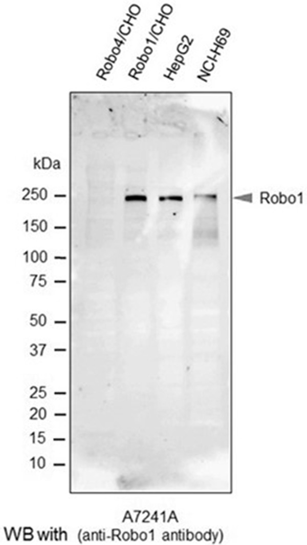
Figure 1.

Western blot analysis with the anti-Robo1 antibody. Robo4/CHO (negative control), Robo1/CHO (positive control), HepG2 and NCI-H69 cells were homogenized in RIPA buffer, and subjected to SDS-PAGE followed by western blot analysis using the A7241A anti-human Robo1 monoclonal antibody. The arrowhead in the image by the ECL method indicates the Robo1 band.
