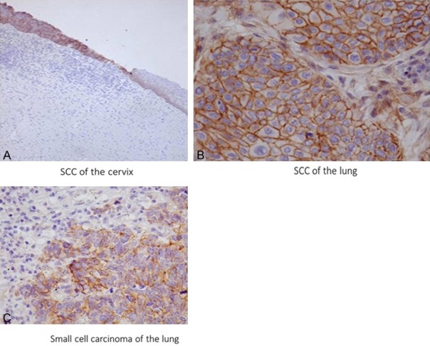
Figure 4.

Positive immunostaining for epithelial cells of squamous cell carcinoma of the uterine cervix (A), squamous carcinoma of the lung (B), and small cell carcinoma of the lung (C). Note that strong membrane staining is apparent in squamous cell carcinoma of the uterus, but not in non-neoplastic squamous epithelium (right) except weakly in the basal layer. Robo1 immunostaining (A) ×100; (B, C) ×400.
