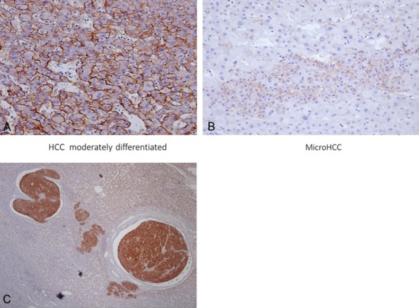
Figure 5.

Robo1 staining in neoplastic and non-neoplastic liver tissues. Normal liver tissue (B, C) lacks staining, in contrast to HCC positivity. HCC exhibits strong membrane expression of Robo1 (A). A small focus of HCC can be seen surrounded by normal liver tissue (B). Tumor cells of HCC invading into vascular space are evident (C). Robo1 immunostaining (A, B) ×200; (C) ×40.
