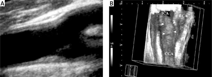
Figure 4.
Seventy-one-year-old woman with asymptomatic carotid artery plaque. A – Hypoechogenic type II atheroma plaque with a smooth surface on the far wall of the common carotid artery on 2D ultrasonographic examination (longitudinal view); B – however, on 3D ultrasonographic imaging, an irregular plaque surface is observed (arrowheads)

