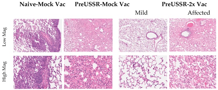
Figure 4.
Histopathological analysis shows differential lung pathology on Day 14 post challenge dependent on influenza virus and vaccination history. Respiratory tissue was collected from ferrets Day 14 post challenge and the lungs were processed for histopathological assessment. Tissue morphology was assessed by hematoxylin & eosin staining. Lungs were analyzed by microscopy from at least three ferrets per group. Images were captured as described and representative images from the groups Naïve-Mock Vac; PreUSSR-Mock Vac; and PreUSSR-2x Vac are shown. High resolution scans were performed using an Aperio ScanScope XT (Leica Biosystems, Concord, Canada) at 40× magnification. Images of the scans were captured using the HALO program from UHN AOMF (Advanced Optical Microscopy Facility, Toronto, Canada) at 5× (low) or 20× (high) magnification of the scan.

