Figure 1.
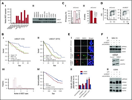
Increased UBE2T expression regulates HR activity in myeloma cells. (A) UBE2T is overexpressed in MM. UBE2T expression evaluated in normal PBMC samples (n = 3) and 8 MM cell lines by real-time qRT-PCR (I) or western blotting (II). (B) UBE2T expression and copy-number correlate with survival in a myeloma dataset (GSE39754; n = 170). Panels I and II show the association of UBE2T expression with overall survival (OS) (I) and event-free survival (EFS) (II). Panels III and IV show UBE2T copy-number alterations (III) and their correlation with overall survival (IV). (C) UBE2T regulates HR in MM cells. (I) HR monitored by the DRGFP assay. MM1S cells chromosomally integrated with the HR repair reporter substrate (DRGFP) were infected with lentiviral shRNA targeting UBE2T, RAD51, or control shRNA. The cells were then infected with a plasmid (AdGNU24i) that expresses the I-SceI endonuclease to initiate HR that is detected by flow cytometry as described in “Methods.” The fraction of GFP-positive cells, representing HR events, for control shRNA was taken as 100%, and other treatments were normalized to that as a percentage. No I-SceI, negative control with no HR; shCONTROL, cells transduced with control shRNA; shRAD51, cells transduced with RAD51 shRNA; shUBE2T, cells transduced with UBE2T shRNA. (II) HR monitored by strand exchange (SE) assay. Control and UBE2T knockdown MM.1S cells were analyzed for homologous strand exchange activity, as described in “Methods.” (D) Impact on cell cycle. Control and UBE2T knockdown MM.1S cells were analyzed for cell cycle using flow cytometry. (E) UBE2T is required for RAD51 focus formation. MM.1S cells transduced with lentivirus expressing the indicated shRNAs were treated with or without CPT (1 μM) for 1 hour and drug washed off. Cells were further cultured in drug-free medium for 6 hours and stained with antibodies to γH2AX and RAD51; DAPI staining was performed to define nuclei. Representative images of γH2AX and RAD51 foci (I) and bar graph showing percentage of cells with ≥5 nuclear γH2AX or RAD51 foci ± standard error of the mean (II) are shown. *P < .05; **P < .01. (F) UBE2T is required for DNA end resection. Myeloma cells, MM.1S (I) or U266 (II) were transduced with control shRNA (shCONTROL) or one targeting UBE2T (shUBE2T) and treated with CPT for 1 hour and evaluated for RPA32 and its phosphorylated form (pRPA S4/8; a marker of DNA end resection). Bottom figure in panel I shows the western blot confirming UBE2T knockdown in MM.1S cells. FITC, fluorescein isothiocyanate; PI, propidium iodide.

