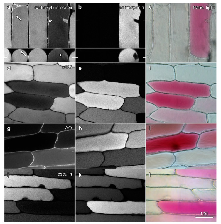
Figure 2.
Fluorescence from dyes in the vacuole is lower in the presence of vacuolar anthocyanin. Onion epidermal peels were loaded with dyes (6 h) and imaged by confocal microscopy, with fluorescence images of the dyes (left column; a,d,g,j) collected concurrently with anthocyanin fluorescence (central column; b,e,h,k) and colour transmitted light images (right column; c,f,i,l). (a–c) Carboxyfluorescein imaged with blue excitation (488 nm), with cytoplasmic fluorescence highlighted in a red cell (asterisk) arrow). The inset shows a single vertical section (XZ) imaging generated at the location marked by bars in (a). (d–f) Monochlorobimane (MCB) imaged with violet excitation (405 nm). (g–i) Acridine orange (AO) imaged with blue excitation (488 nm). (j–l) Esculin imaged with violet excitation (405 nm). Bar in (i) = 100 µm for all images.

