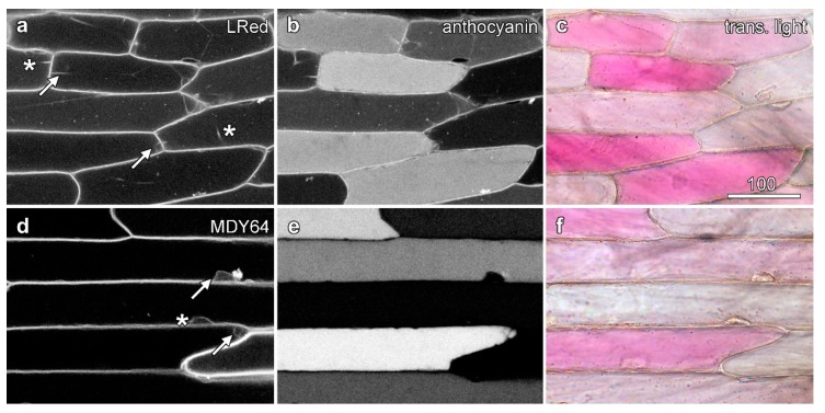
Figure 3.
Tonoplast labelling is unaffected by the presence of anthocyanin. Epidermal peels loaded with dyes were imaged with sequential scanning. Images are confocal optical sections of dye fluorescence (left column; a,d), with the tonoplast labelling in anthocyanic cells indicated with arrows and in white cells with asterisks, anthocyanin fluorescence (central column; b,e), and a colour transmitted light image (right column; c,f). (a–c) Lysotracker Red DND-99 (LRed) imaged with green excitation collecting shorter wavelength red fluorescence, with the longer wavelength red fluorescence collected from anthocyanin with some bleed-through of the Lysotracker. (d–f) MDY-64 imaged with blue (473 nm) excitation. Bar in (a) = 100 µm for all images.

