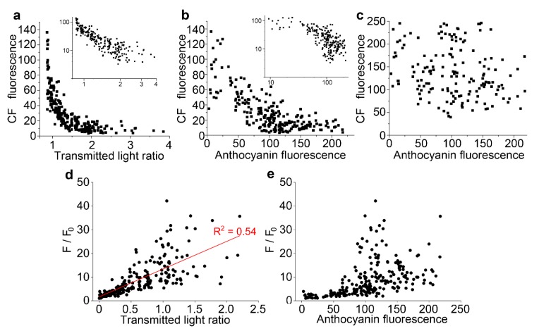
Figure 5.
Vacuolar fluorescence of carboxyfluorescein is lower in the presence of anthocyanin. For individual cells from multiple epidermal peels in a single representative experiment, anthocyanin and carboxyfluorescein fluorescence were measured in arbitrary units, with anthocyanin also measured as a transmitted light ratio, the image intensity at 633 nm divided by the intensity at 561 nm. (a) Vacuolar carboxyfluorescein showed a strong negative correlation with the transmitted light ratio. The inset shows the same dataset replotted on a double-log plot. (b) Vacuolar carboxyfluorescein showed a negative correlation to anthocyanin fluorescence. The inset shows the same dataset replotted on a double-log plot. (c) Cytoplasmic carboxyfluorescein did not correlate to anthocyanin fluorescence. (d,e) Stern–Volmer plots, used to investigate the quenching of fluorescence, correlate maximum dye fluorescence divided by the dye fluorescence (F/F0) against anthocyanin concentration: when plotted against transmitted light ration (d), a straight line was observed whereas plotting against anthocyanin fluorescence did not give a straight line (e).

