Figure 3.

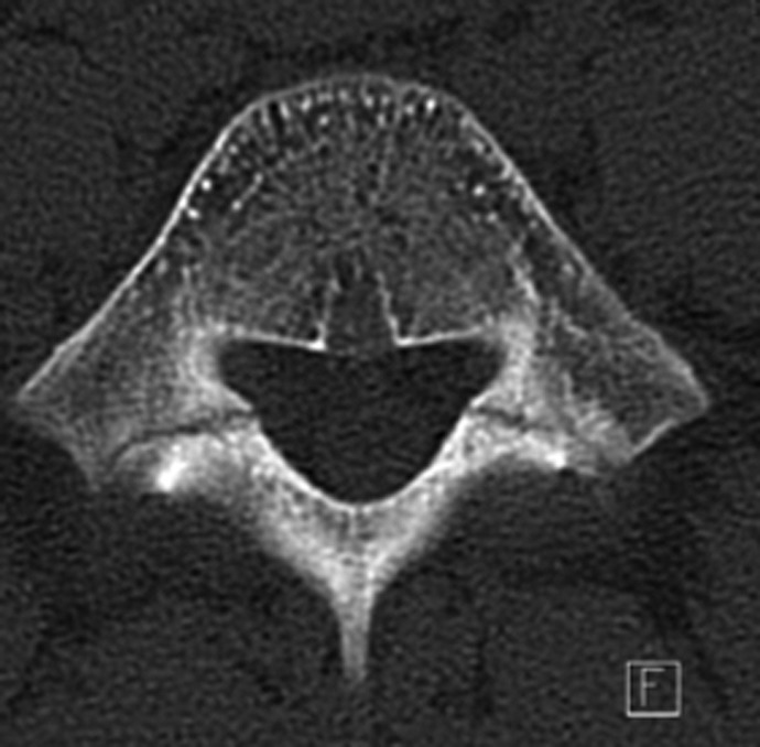
X-ray of the lumbar spine (axial view) showing bilateral spondylolytic defects. This radiograph is from one of the author’s patients.

X-ray of the lumbar spine (axial view) showing bilateral spondylolytic defects. This radiograph is from one of the author’s patients.