Figure 3.
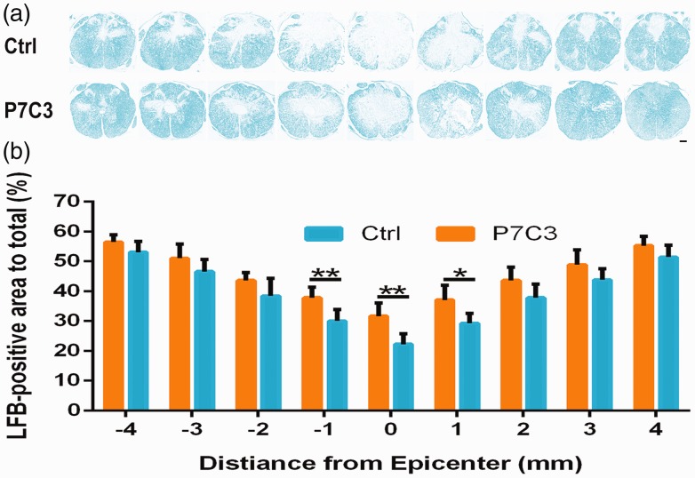
Treatment with P7C3 increases residual myelination following SCI. (a) Representative images of white matter from the lesion epicenter (0) and at 1, 2, 3, and 4 mm rostral (+) and caudal (−) to the epicenter, as detected by LFB staining. (b) Quantification of LFB-stained areas at various distances from the lesion epicenter. Data represent mean ± SD (n = 6). *P < 0.05; **P < 0.01. Scale bar = 200 μm. (A color version of this figure is available in the online journal.)

