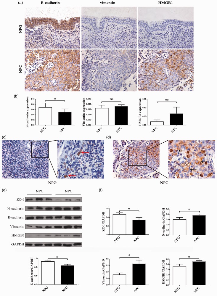
Figure 1.
Expression of EMT markers and HMGB1 in non-keratosis NPC and NPG tissues. (a) An immunohistochemistry assay was performed to detect the staining of E-cadherin, vimentin, and HMGB1 in non-keratosis NPC and NPG tissues (×400). (b) The expression of E-cadherin, vimentin and HMGB1 in non-keratosis NPC and NPG tissues was determined by immunohistochemical semiquantification. Data are presented as mean ± SD. *P < 0.05, **P < 0.01. (NPC, n = 10 NPG, n = 10). (c, d) The location of HMGB1 in non-keratosis NPC and NPG tissues (×400, ×1000). The arrows show the location of HMGB1. (e) Protein expressions of ZO-1, N-cadherin, E-cadherin, vimentin, and HMGB1 were examined by Western blot. The experiment was performed in triplicate. (f) Quantitative analyses of EMT markers and HMGB1. Data are expressed as mean ± SD. *P < 0.05. The data represent the average of three experiments. (A color version of this figure is available in the online journal.)
NPC: nasopharyngeal carcinoma; NPG: nasopharyngitis; GAPDH: glyceraldehyde 3-phosphate dehydrogenase; HMGB1: high mobility group box 1.

