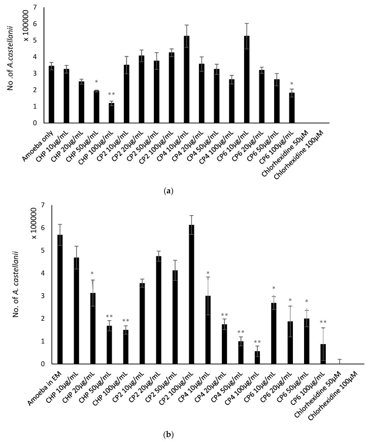
Figure 7.
(a) Effects of different sizes of cobalt nanoparticles (CHP, CP2, CP4, and CP6) on encystation of A. castellanii at concentrations of 10, 20, 50, and 100 µg/mL after 72 h at 30 °C. A. castellanii trophozoites were incubated with chlorhexidine (positive control) and amoeba alone in phosphate-buffered saline (PBS) with encystation medium (EM), 50 mM MgCl2, and 10% glucose) as a negative control. After 72 h, the number of mature cysts were counted using a hemocytometer, and 0.1% sodium dodecyl sulphate (SDS) was used to dissolve remaining trophozoites. The results obtained represent the mean and standard error of three independent reproducible experiments performed in duplicate. * P < 0.05, ** P < 0.01 and *** P < 0.001, using a two-sample t-test and two-tailed distribution. (b) The effects of different sizes of cobalt nanoparticles (CHP, CP2, CP4, and CP6) on excystation of A. castellanii at concentrations of 10, 20, 50, and 100 µg/mL at 72 h and 30 °C. A. castellanii cysts were incubated with chlorhexidine (positive control) and amoeba alone (negative control). After 72 h, the number of viable trophozoites was enumerated by Trypan blue exclusion assay. The results obtained represent the mean and standard error of three independent reproducible experiments performed in duplicate. * P < 0.05, ** P < 0.01 and *** P <0.001, using a two-sample t-test and two-tailed distribution.

