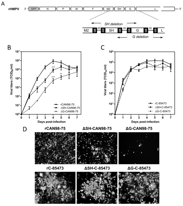
Figure 1.
Construction of recombinant human metapneumovirus (HMPV) viruses and in vitro replicative capacity of SH and G-deleted rHMPV viruses. (A) Schematic representation of the genome of a recombinant GFP-expressing recombinant HMPV (rHMPV) viruses, with specific focus on the region of the genome describing the position of primers used for the deletion strategy (arrow, Table 1). GS: gene start. GE: gene end. IG: intergenic sequence. (B,C) LLC-MK2 monolayers in 24 wells-plates were infected with each of the six recovered rHMPV strains (rCAN98-75, ΔSH-CAN98-75, ΔG-CAN98-75; rC-85473, ΔSH-C-85473, ΔG-C-85473) at a MOI of 0.01. Supernatants were harvested every 24 h for seven days, frozen, sonicated and titrated as tissue culture infectious doses (TCID50)/mL on LLC-MK2 cells. Growth curves represent mean titers of two independent experiments, with each time point titrated in triplicate. ** p < 0.01, *** p < 0.001 when comparing each ΔSH/ΔG virus to its wild type (WT) counterpart using repeated measures two-way ANOVA. (D) Images of representative cytopathic effects and spread of each virus at three days post-infection were captured using fluorescent microscopy (10 × magnification).

